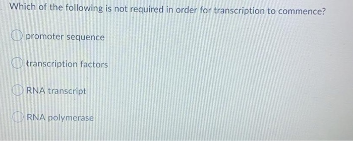
Which test is used to diagnose conditions associated with abnormal bleeding and to monitor anticoagulant therapy? This chapter discusses the new developments in cardiac pet tracers, cyclotrons, and delivery systems.
Which Imaging System Combines Tomography With Radionuclide Tracers. And combines with a negative electron. The scanning tests use a special camera to take pictures of certain tissues in the body for clinical diagnosis after a radioactive tracer (radiopharmaceutical) is given to the patient. Clinical spect imaging systems typically have two or three planar scintigraphy cameras that rotate around the patient. The field of cardiovascular pet imaging is constantly evolving, and this includes all aspects of tracers, production of tracers, and delivery to the patient.
 Combining External Beam Radiation And Radionuclide Therapies: Rationale, Radiobiology, Results And Roadblocks - Clinical Oncology From clinicaloncologyonline.net
Related Post Combining External Beam Radiation And Radionuclide Therapies: Rationale, Radiobiology, Results And Roadblocks - Clinical Oncology :
After injection of the chosen radiotracer, the isotope is extracted from the blood by viable myocytes and retained within the myocyte for some time. Study of the nature, uses, and effects of drugs for medical purposes. Which imaging system combines tomography with radionuclide tracers to produce enhanced images of selected body organs or areas? Spect imaging is similar to pet in that both require a radiotracer to form the image.
Routinely in radionuclide bone imaging for malignancy
Positron emission tomography (pet) is a functional imaging technique that uses radioactive substances known as radiotracers to visualize and measure changes in metabolic processes, and in other physiological activities including blood flow, regional chemical composition, and absorption. The positron and electron are annihilated, which. Routinely in radionuclide bone imaging for malignancy This review provides an overview of the development of positron emission tomography (pet) radiotracers for in vivo imaging of ar system in the brain. Radionuclide & molecular imaging is one of the common medical detection methods these days, which diagnose and cure diseases using radiopharmaceuticals. _____ (pet) combines tomography with radionuclide tracers to produce enhanced images of selected body organs or areas.
 Source: coursehero.com
Routinely in radionuclide bone imaging for malignancy And combines with a negative electron. Which imaging system combines tomography with radionuclide tracers to produce enhanced images of selected body organs or areas?
 New Radionuclides And Technological Advances In Spect And Pet Scanners") Source: researchgate.net
Source: researchgate.net
It is very similar to conventional nuclear medicine planar imaging using a gamma camera, but is able to provide true 3d information. It is very similar to conventional nuclear medicine planar imaging using a gamma camera, but is able to provide true 3d information. Positron emission tomography also known as pet imaging, combines tomography with radionuclide tracers to produce enhanced images of selected body organs otc drug
 Source: slideplayer.com
(1 point) magnetic resonance imaging. The 3d images are computer generated from a large number of projection images of the body recorded at different angles. This chapter discusses the new developments in cardiac pet tracers, cyclotrons, and delivery systems.
 Source: coursehero.com
Spect imaging is similar to pet in that both require a radiotracer to form the image. Combines tomography with radionuclide tracers to produce enhanced images of selected body organs or areas: It is very similar to conventional nuclear medicine planar imaging using a gamma camera, but is able to provide true 3d information.
 Source: chemistry-europe.onlinelibrary.wiley.com
The 3d images are computer generated from a large number of projection images of the body recorded at different angles. After injection of the chosen radiotracer, the isotope is extracted from the blood by viable myocytes and retained within the myocyte for some time. This review provides an overview of the development of positron emission tomography (pet) radiotracers for in vivo imaging of ar system in the brain.
 Source: mdpi.com
Molecular radiotherapy combines the potential of a specific tracer (vector) targeting tumor cells with local radiotoxicity. After injection of the chosen radiotracer, the isotope is extracted from the blood by viable myocytes and retained within the myocyte for some time. _____ (pet) combines tomography with radionuclide tracers to produce enhanced images of selected body organs or areas.
 Source: jnanobiotechnology.biomedcentral.com
And combines with a negative electron. Positron emission tomography (pet imaging) definition. Combined with tracer distribution imaging (fluorescence and/or nuclear).
 Source: coursehero.com
Positron emission tomography also known as pet imaging, combines tomography with radionuclide tracers to produce enhanced images of selected body organs otc drug Combined with tracer distribution imaging (fluorescence and/or nuclear). It is very similar to conventional nuclear medicine planar imaging using a gamma camera, but is able to provide true 3d information.
 Pitfalls And Limitations Of Radionuclide Planar And Hybrid Bone Imaging") Source: researchgate.net
Source: researchgate.net
The radionuclides used in spect imaging typically emit a single gamma photon as a component of their disintegration. Positron emission tomography (pet imaging) definition. _____ (pet) combines tomography with radionuclide tracers to produce enhanced images of selected body organs or areas.
 First-In-Class Positron Emission Tomography Tracer For The Glucagon Receptor") Source: researchgate.net
Source: researchgate.net
After injection of the chosen radiotracer, the isotope is extracted from the blood by viable myocytes and retained within the myocyte for some time. This review provides an overview of the development of positron emission tomography (pet) radiotracers for in vivo imaging of ar system in the brain. Different tracers are used for various imaging purposes, depending on the target.
Source: clinicaloncologyonline.net
Spect imaging is similar to pet in that both require a radiotracer to form the image. Positron emission tomography also known as pet imaging, combines tomography with radionuclide tracers to produce enhanced images of selected body organs otc drug Which test is used to diagnose conditions associated with abnormal bleeding and to monitor anticoagulant therapy?
 Source: nature.com
This chapter discusses the new developments in cardiac pet tracers, cyclotrons, and delivery systems. Radionuclide & molecular imaging is one of the common medical detection methods these days, which diagnose and cure diseases using radiopharmaceuticals. And combines with a negative electron.
 Source: thno.org
The scanning tests use a special camera to take pictures of certain tissues in the body for clinical diagnosis after a radioactive tracer (radiopharmaceutical) is given to the patient. Spect imaging is similar to pet in that both require a radiotracer to form the image. (1 point) magnetic resonance imaging.
 Source: coursehero.com
The radionuclides used in spect imaging typically emit a single gamma photon as a component of their disintegration. And combines with a negative electron. The scanning tests use a special camera to take pictures of certain tissues in the body for clinical diagnosis after a radioactive tracer (radiopharmaceutical) is given to the patient.
 Source: thno.org
The positron and electron are annihilated, which. This review provides an overview of the development of positron emission tomography (pet) radiotracers for in vivo imaging of ar system in the brain. Study of the nature, uses, and effects of drugs for medical purposes.
 Source: jnanobiotechnology.biomedcentral.com
The positron and electron are annihilated, which. Positron emission tomography also known as pet imaging, combines tomography with radionuclide tracers to produce enhanced images of selected body organs otc drug This review provides an overview of the development of positron emission tomography (pet) radiotracers for in vivo imaging of ar system in the brain.
 Source: sciencedirect.com
_____ (pet) combines tomography with radionuclide tracers to produce enhanced images of selected body organs or areas. Routinely in radionuclide bone imaging for malignancy Several imaging systems which combine ßuorescence and radionuclide have been developed in recent years.
 Source: coursehero.com
Combined with tracer distribution imaging (fluorescence and/or nuclear). This review provides an overview of the development of positron emission tomography (pet) radiotracers for in vivo imaging of ar system in the brain. Radionuclide & molecular imaging is one of the common medical detection methods these days, which diagnose and cure diseases using radiopharmaceuticals.
 Source: slideplayer.com
Spect imaging is similar to pet in that both require a radiotracer to form the image. It is very similar to conventional nuclear medicine planar imaging using a gamma camera, but is able to provide true 3d information. Routinely in radionuclide bone imaging for malignancy
 Source: coursehero.com
The field of cardiovascular pet imaging is constantly evolving, and this includes all aspects of tracers, production of tracers, and delivery to the patient. Molecular radiotherapy combines the potential of a specific tracer (vector) targeting tumor cells with local radiotoxicity. Study of the nature, uses, and effects of drugs for medical purposes.
Also Read :